
Effortless Removal: Laser Skin Tag Removal for Smooth & Clear Skin!
OVERVIEW
skin growth is a benign condition which consists of extra unwanted tissue protruding out of skin or on skin. This are of various types:
SKIN TAGS
Skin tags are noncancerous growth which are small and same color to the skin. Mostly they are seen in clusters and typically present where skin rubs to each other. It irritates skin and its appearance bothers a lot.
It doesn’t affect health or its not harmful. But If present in neck or face areas its worrying due to its look. Mostly it causes discomfort due to friction while wearing clothes or jewelry. Very rare cases it leads to bleeding.
The causes are not exactly known but most of the researchers said its hereditary or mostly seen who are having issues like diabetes, obesity or hormonal changes.
This can grow in any are but mostly it is seen in armpits, eyelids, groin, thighs, neck, under breast or genitals.
There are lot of over the counter products sold in market for skin tag removal but its use at home is not safe as it has a risk of bleeding, scar mark, infection, incomplete removable.
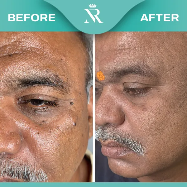
Our dermatologists firstly examine the skin tag and then after some pathological investigations are normal patient is given appointment for skin tag removal. Local anesthesia is given with minute syringe which allows procedure to done painless. RF CAUTERIZATION is done which removes skin tag from root level so that it should not grow again and remains scar less. Our doctors are skilled to remove skin tags from eyelid also. Topical antibiotic is advised to apply. Can resume work immediately without any restrictions.
Maintain healthy diet with regular exercise can avoid skin tags.
Cost of skin tag removal is affordable and time procedure. It costs from rs. 10/- to rs.20/-.
KELOIDS
- Overview
Keloid is scar growth with fibrous tissue over the skin. It comes from overgrowth of scar tissue. It causes cosmetic concern rather than to health. It grows whenever there is skin injury but mostly seen on chest, shoulders, cheeks, earlobes.
- Its thick, irregular, lumpy.
- It causes itchiness and irritation.
- Its color is lighter and shiner to skin color.
Mainly seen in area wherever there is wound but mostly present in middle chest area, shoulders, cheek or earlobes.
It is hypertrophied scar which comes post wound. It grows whenever there is wound may be due to injury, insect bite, injection, post-surgery, burns, scratches, etc.
Its wound healing dysfunction where excess collagen produces more tissue. Its non-cancerous and may fade on its own in some cases. Mostly it is inherited. And one has having one keloid may have chances of future growth in other areas.
- Avoid injury.
- If there is surgery one should inform his doctor about his tendency to develop keloid post injury.
- If there is wound he should be precautious during wound healing process and follow strict aseptic care.
- Topical creams: corticosteroids are advised to avoid itching and irritation.
- Corticosteroid injection: mostly it gets flatten with 4-6 sessions of injection in keloid when its administered in early stages and if its small.
- Surgical removal: mostly surgical removal is quite reversible but can try if present on prominent areas.
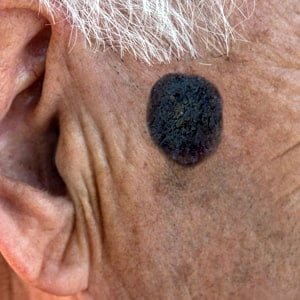
MOLES
- Overview
This is very common skin growth which is mostly harmless and sometimes if on face known as beauty mark. Still one should not ignore if one feels something abnormal with it and consult dermatologist. They appear as small, dark brown spots caused by cluster of pigment forming cells i.e. melanocytes.
- Symptoms of mole
- Mostly they appear in early childhood or till 20s. often some hair develop from mole. Its usually brown spot. This can appear in scalp, anywhere on skin, armpits, between fingers or toes.
- Color
- May vary to black, blue, red or pink also.
- Shape
- Is oval or round
- Size
- Is not more than 6mm in diameter
WHEN MOLE IS A CONCERN?
The ABCDEs are important signs of moles that could be cancerous. If a mole presents any of the signs listed below, one should seek attention and consult dermatologist:
Asymmetry
Half side of mole if doesn’t match to another side of mole.
Border
If the edges of your mole are irregular.
Color
If the color of your single mole is not the same throughout, or if has shades of multiple colors such as tan, brown, black, blue, white, or red.
Diameter
If the diameter of your mole is larger than the eraser of a pencil.
Elevation/Evolution
If your mole shows growth, or it changes over a short period of time.
The most common location for melanoma in men is the back; in women, it is the lower leg. Melanoma is the most common cancer in women ages 25 to 29.
After consultation with dermatologist he will advice skin biopsy and if test is positive one need to be immediately go for complete excision of mole.
TYPES OF MOLE
Congenital mole
Congenital nevi occurs from birth itself, this are more likely to turn out in melanoma.
Dysplastic mole
This are larger in size more than 0.8mm and irregular shaped. These moles have history of inheritance and ae more likely to be malignant melanoma. They appear uneven with dark brown center and lighter edge.
Common mole
This are with uniform edge and doesn’t grow in size and remains same for years.
CAUSES OF MOLE
When pigment cells in skin called melanocytes grow in clusters they are referred to as moles. This are mostly inheritance. And cause remains unknown. Mostly seen in tropical region or exposure to UV rays.
- How do I check for moles
- One should protect his skin if he has moles by following measures:
- Protect skin from UV rays.
- Avoid too sun exposure, sun protective measures.
- Use sun screen all times.
- Use sunglasses, long sleeve clothes, hats, etc.
- Avoid tanning lamps or lights having UV rays.
TREATMENT OF MOLES
Moles can be easily diagnosed by dermatologist and easily removable without any discomfort. It is very short time procedure and and as outpatient basis. Moles can be removed as per their condition. Unless moles if bothersome or unattractive to the patient No need of removal. If a dermatologist suggests if seen suspicious for any skin cancer then cant be ignored. Superficial moles can be treated with lasers and with the help of Surgical cautery. Moles which are deeper may require surgical excision with local anaesthesia.
New try to remove mole at home:
One may disfigure your skin, causing a scar or an infection if tried to remove at home with traditional techniques.
MOLLUSCUM CONTAGIOSUM
Overview
Molluscum contagiosum is very common and contagious, viral skin infection. It causes raised, people like noodles or papules on the skin. The papules are generally painless and they do not eat. They are specially present on the trunk of body, arms and legs.
Symptoms
After initial infection, it takes up to 7 days to 6 months for symptoms to arrive. The papules which appear are small, firm, flesh coloured, dome shaped, pearly, wart like spots on the skin. It’s typically 1-5 mm in diameter with a dimpled Centre. Some mollusca have a small white dot with pus and secrets a thick white fluid, if bursted Dimple part me bleed.
Causes
It gets infected, mostly when the immune system is compromised. It spreads through direct contact with an infected person or skin to skin contact or touching the contaminated objects like towels, etc.
Treatment
Its treated with the help of laser therapy or diathermy cautery.
KERATOSIS
Overview
Seborrheic keratosis is a benign lesion on the skin caused by cumulative effects of long-term exposure to the UV light . It is common in elderly or after middle age. It is usually light tan to brown or black.
When should one attend to dermatologist ?
Seborrheic keratosis is harmless. Most of them do not need any treatment, but if any patient gets irritated due to friction on clothing or jewellery or seems to be ugky appearance to the patient, then dermatologist can help to remove it.
Signs of seborrheic keratosis
They usually appear on back or chest or other parts exposed to sun. It grows slowly in group or singly. It is light tan to brown or black with rough texture with bumpy surface. Feel on touch is smooth & greasy. It can grow till 3 cms in diameter.
Who are most likely prone to seborrheic keratosis
People with fair skin, people who get sunburn easily, with sensitive skin, long-term sun, exposure, who play outdoor sports, people who have family history are most likely to have seborrheic keratosis.
How to get rid of seborrheic keratosis
If the lesions are small and in multiple areas then chemical peeling is the best option to exfoliate the upper layer of skin. If more deeper or localised, then can go with by surgical cautery or laser treatments.
How to prevent seborrheic keratosis?
Avoid long-term sun, exposure, habit of applying sunscreen of SPF more than 30, long sleeve shirt and broad brimmed hat if one engaged in long outdoor activities is always helpful.
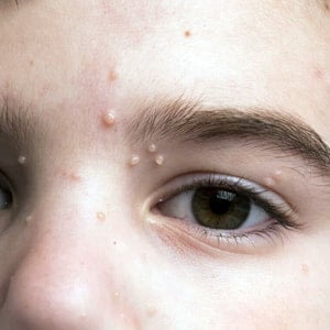
WARTS
Overview
- - Warts are infections in the skin caused by human papilloma virus (HPV).
- - Warts are common in childhood till 12 to 6 years.
- - Warts are easily treatable.
Symptoms of warts
- - Words are mostly affected on face, feet, knees and hands.
- - Warts may occur singly or in clusters.
- - These have a rough or smooth surface and average size ranges from 1 to 10 mm only.
- - These are small, raised on the skin.
TYPES OF WARTS
These appear like hard, raised lumps with rough surfaces. Common sites are knees and hands, but can occur at any part of the body.
These appear smooth, flattened lumps. Can occur at any part but most commonly on face, lower legs & hands are common sites.
These appears on hands & soles in clusters.
These appears usually on soles of feet which look like small, hard bumps and having tiny black dots on them.
It has grainy cauliflower appearance which are sexually transmitted. Penis, vulva & anus are mostly affected . It may be initial sign or predispose to cancer. Patients should not ignore and should immediately consult a doctor.
RISK FACTORS OF WARTS WHICH NEEDS QUICK ATTENTION
Anyone can have warts but the factors which should not be ignored are as follows:
- Injuries to skin
- Skin infections which cracks skin
- Frequent wetting of hands
- Hands or feets sweating heavily
- Who have habit of nail biting & swimming regularly in public swimming pools.
- Direct contact with the person already infected
- Itching of the warts
WARTS TREATMENT
Topical agents
Creams containing salicylic acid or lactic acid are the best help initially.
Immune system stimulator
To develop the immune system to fight viral infection
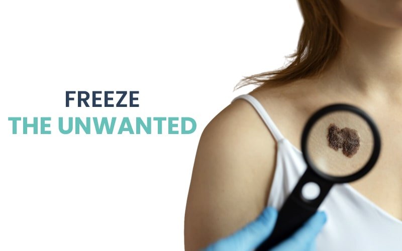
Cryotherapy
Words or frozen with liquid nitrogen to get rid of warts
Electric cautery
It helps to remove from base and heals within a week.
Laser therapy
iIhelps to remove superficial warts.
Immunisation
To the children aged to 12 to 25 years who are at risk.
RELATED VIDEO
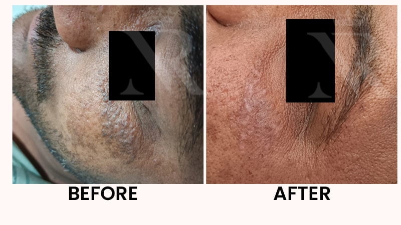
Before & After